ASA for LiteScope AFM
The Alemnis nanoindenter module (ASA) can be used as an optional accessory for the atomic force microscope (AFM) LiteScope, designed for integration into scanning electron microscopes.
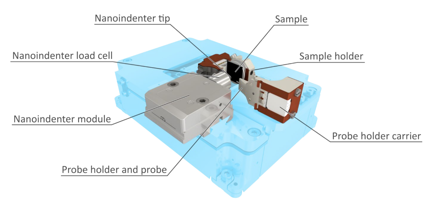
The resulting combination of three complementary techniques enables micromechanical experiments to be performed while observing the specimen with superb SEM magnification and analysing the indented specimen with sub-nanometer resolution using LiteScope. This unique solution is designed for maximum versatility and enables a wide range of novel and complex applications.
Added Values
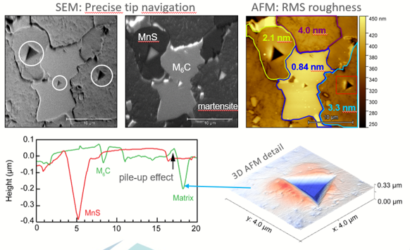
- Quantitative analysis of mechanical properties (hardness, Young’s modulus, activation volume).
- Precise analysis of indent topography and pre-indented sample roughness.
- Utilization of SEM and AFM analysis for optimal spot selection for nanoindentation.
Request a quote for Alemnis Standard Assembly.
Quote